
synECi18

What is Synthesized ECG?
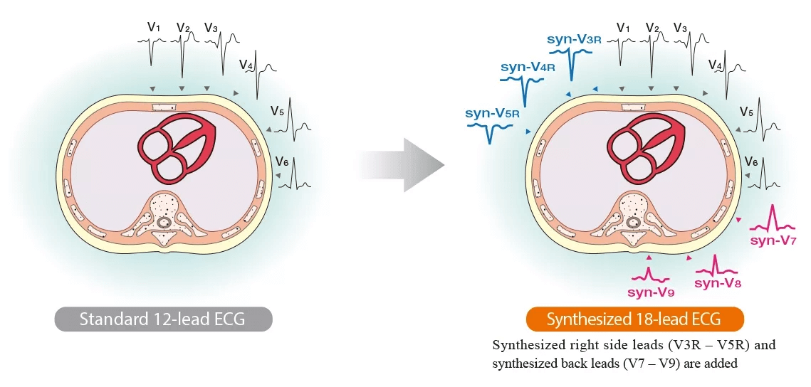
The most common ECG exam is the standard 12-lead ECG. It is simple to measure, has a low burden on the body, and observing the heart from these 12 directions provides a lot of information with a wide range of clinical applications. However, some areas, especially pathological changes in the right ventricle and the posterior wall, cannot be observed from the 12-lead ECG.
Synthesized 18-lead ECG uses the 12-lead ECG waveforms to mathematically derive the waveforms of the right chest leads (V3R, V4R, V5R) and back leads (V7, V8, V9). The measurement procedure is the same as the standard 12-lead ECG, but more information can be obtained. 18-lead synthesized ECG is expected to be useful in detecting right side and posterior infarction.

Principle of Synthesized Waveforms
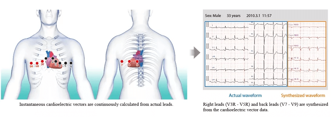
Instantaneous cardioelectricvectors are continuously measured from the standard 12-lead ECG data, and the ECG for the right leads (V3R, V4R and V5R) and back leads (V7, V8 and V9) is synthesized from this data.


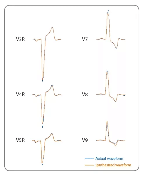
The example on the left shows actually measured waveforms and synthesized waveforms. Other data also has good correlation with actually measured ECG. This suggests that we can obtain useful information which corresponds to the condition of the heart.
References
1. US Patent No. : US8.005.532: Electrocardiograph with extended function, and extended lead electrocardiogram deriving method.Inventor: DamingWei. ph. D
2. Wei D., et al.: Derived electrocardiograms on the posterior leads from 12-lead system: method and evaluation.Proceedings of the 25th Annual International Conference of IEEE IEMBS 2003; 1: 74-77.
3. KatohT., et al., Clinical Significance of Synthesized Posterior Right-Sided Chest Lead Electrocardiograms in Patients with Acute Chest Pain. J Nippon Med Sch 2011: 78 (1): 22-29.
4. Kenichi Harumi. Guide for electrocardiographic diagnosis. Sogo Rinsho. extra number. 1995.
5. WlliottM Antman. MD. FACC. FAHA. Chalret al. ACC/AHA Guidelines for the Management of Patients With ST-Elevation Myocardial Infarction. Circulation. 2004:110:e82-e292.
6. The 73thAnnual Scientific Meeting of the Japanese Circulation Society, March 20-22, 2009Diagnostic Improvement for Detecting Posterior Myocardial Infarction Using Lead V9 on Surface ECG: Comparison with Echocardiography.Nobuhiko Haruki*, Hidetoshi Yoshitani*, Hiromi Nakai*, Kyoko Otani*, Kyoko Kaku*, Toshiyuki Ota*, Yutaka Otsuji*
7. International Society for Holter and Noninvasive Electrocardiology(ISHNE)2009June 4-62009YokohamaDiagnostic Improvement for Detecting Posterior Myocardial Infarction Using Virtual ECG: Comparison with Echocardiography.Nobuhiko Haruki*1, Masaki Takeuchi*1, Hiromi Nakai*1, Kyoko Kaku*1, Hidetoshi Yoshitani*1, Jiro Suto*2, Yutaka Otsuji*1*1Second Department of Internal Medicine, University of Occupational and Environmental Health, School of Medicine*2NIHON KOHDEN CORPORATION, Japan
8. The 29th Annual Meeting of the Japanese Society of Electrocardiology, September 18-22, 2011Pitfall of Synthesized Posterior/Right-Sided Chest Lead Electrocardiograms.Yukio Saitoh*, Yoshinari Goseki*, YoshinaoYazaki*, Akira Yamashina** The Second Department of Internal Medicine, Tokyo Medical University, JapanP-Wave Dispersion in Synthesized 18-Lead ECG of Paroxysmal Atrial Fibrillation Patients.YasunoriHiranuma*, MarehikoUeda*, TakatsuguKajiyama*, NaotakaHashiguchi*, Tomonori Kanaeda*, Yusuke Kondo*, Masahiro Nakano*, Yoshio Kobayashi** Department of Cardiovascular Science and Medicine, Chiba University Graduate School of Medicine, Japan
9. About the Normal Range in the Addition Leads(V3R,V4R,V5R, V7,V8,V9) When We Used Synthesized 18 Leads ECG.Jiro Suto*1, Tsuneo Takayanagi*1, Akira Yamashina*2*1Engineering Depertment1 Biomedical Instrument Technology Center NIHON KOHDEN CORPORATION, Japan*2Cardiovascular Internal Medicine, Tokyo Medical University Hospital, Japan