Cardiac and respiratory monitoring are standard practices in the intensive care unit (ICU) environment today, however, electroencephalography (EEG) which plays a crucial role in determining the current status of the patient is not. Despite challenges associated with continuous EEG monitoring (cEEG) in ICU, during this crucial phase of patient care, Nihon Kohden offers multiple solutions for cEEG measurement.
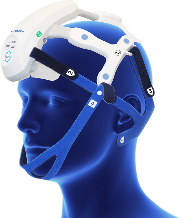
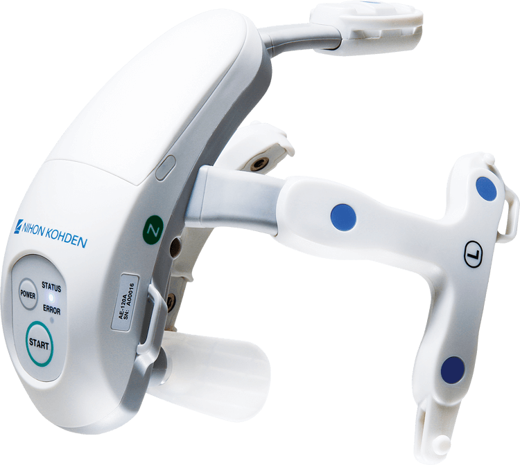
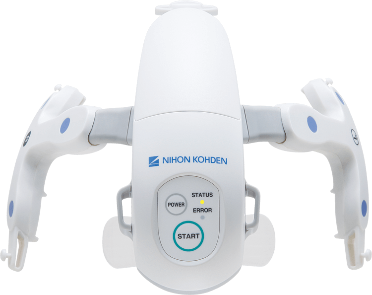
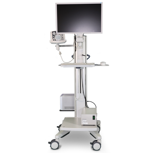
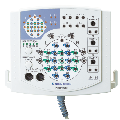
Our innovative wireless EEG headset (CerebAir) enables the streamlined preparation and measurement enable faster EEG acquisition and long-term monitoring. Clinicians are better supported with seizure detection algorithms enabled in the EEG trend software with the wired 38-channel junction box, allowing for the quick identification of seizure activity resulting in quicker clinical intervention for the patient. Simplified EEG monitoring is available with modules on Nihon Kohden Patient Monitors, allowing for the EEG data to be displayed on the central nursing station.
As a result, continuous EEG monitoring in ICU can be become a standard parameter for comprehensive patient care in this critical care setting.
")