Image
hero-capone

cap-ONE

Image
cap-ONE logo

What is cap-ONE?

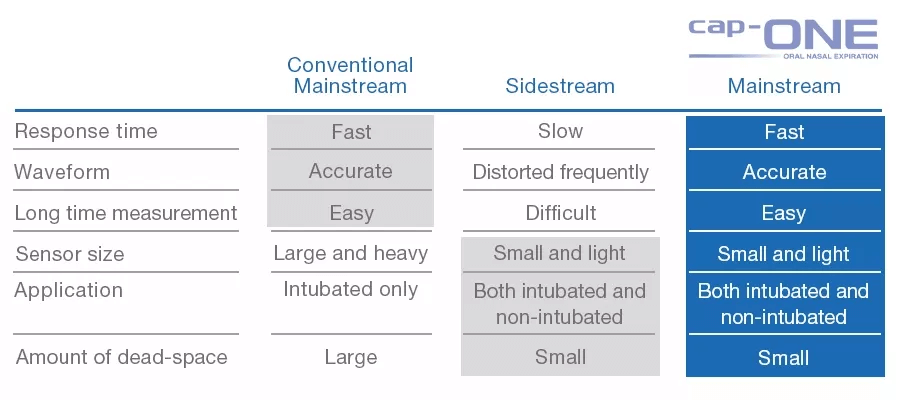
cap-ONE is Nihon Kohden's unique mainstream CO2 sensor for both intubated and non-intubated patients. The ultra compact sensor and unique adapter provide accurate measurements and reduce the burden on the patients. 
cap-ONE overcomes the disadvantages and maintains all the advantages of mainstream sensor for optimal capnography.

Image
Image tech cap-ONE 22

Features

Heaterless and Motorless structure

Our CO2 sensor is ultra-compact weighing only 4 g, with a heaterless and motorless structure, that meets the United States Military Standard* for shock resistance.


*United States Military Standard: a performance test standard for products used by the US military. This is one of the evaluation standards for products in harsh environments.

Our two mainstream CO2 sensors

Based on the principle of infrared absorption spectroscopy, Nihon Kohden provides two mainstream CO2 sensors: Quantitative method and Semi-quantitative method.


Why is CO2 monitoring important?

CO2 measurement can predict hyperventilation or hypoventilation which can be caused by inadequate analgesia, sedation or other potentially life-threatening causes.

Recommendation from guidelines and studies

Ensure quality of CPR and early indicator for ROSC1

  • A value of less than 10 mmHg is associated with failure to achieve ROSC and may indicate that the quality of chest compressions should be improved

Detect ventilator troubles

  • 8-25% of anesthesia accidents reported as breathing circuit problems
  • Breakdown of breathing circuit problems: Circuit disconnection 8-15%, Misconnection of circuits 2-4%, Displacement of endotracheal tube 2-6%

Avoid unrecognized esophageal intubation2

  • Patients worldwide die from unrecognized esophageal intubation every year, which is an avoidable complication of airway management usually resulting from human error.
  • Checking correct tracheal tube placement using capnography for identification and monitoring of expired carbon dioxide (CO2) is necessary to avoid human error.

Reduce frequency of hypoxemic events3, 4

  • Continual monitoring of ventilatory function with capnography to supplement standard monitoring by observation and pulse oximetry is recommended to address moderate procedural sedation and analgesia
  • Access to capnography provides fewer but more timely interventions for hypoventilation, which leads to fewer episodes of hypoventilation and of oxygen desaturation

Assist in better patient cares and outcomes5

  • The monitoring of ventilation using capnography is vital to titrating sedatives for which different patients have variable levels of sensitivity, which provides early warning signs of adverse respiratory events


1 Part 3: Adult Basic and Advanced Life Support: 2020 American Heart Association Guidelines for Cardiopulmonary Resuscitation and Emergency Cardiovascular Care | Circulation (ahajournals.org)            
2 Paul A. Baker Unrecognisedoesophagealintubation: time for action –ScienceDirect           
3 Practice Guidelines for Moderate Procedural Sedation and Analgesia 2018 | Anesthesiology | American Society of Anesthesiologists (asahq.org)            
4 Langhan, M. L., Shabanova, V., Li, F. Y., Bernstein, S. L., & Shapiro, E. D. (2015). A randomized controlled trial of capnography during sedation in a pediatric emergency setting.The American journal of emergency medicine,33(1), 25–30.           
5 Use of Capnography during Moderate Sedation by Non-Anesthesia Personnel in Various Clinical Settings -Anesthesia Patient Safety Foundation (apsf.org)


Our two mainstream sensors

Quantitative method

Image
Technology CO2 image 01

In the quantitative method, the infrared light transmitted through the airway adapter is split into two by a half mirror. One of which is detected as a signal output (Vs), the other passes through the gas cell* and is detected by another photo detector as the standard output (Vr). The partial pressure is calculated from the ratio of this signal output Vs and standard output Vr. *gas cell: a cell containing 100% CO2 gas

Image
image tech co2 02
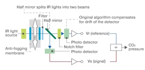
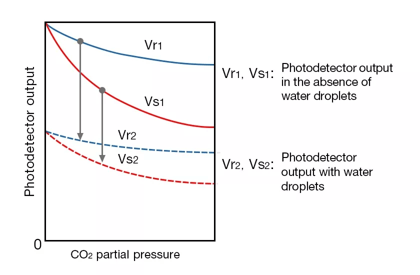
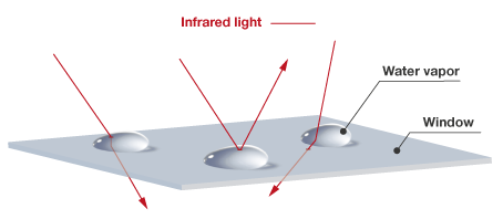
In the quantitative method, even the light intensity decreases, the ratio of signals and standard output (Vs/Vr) won’t change. Therefore, it is less likely to be affected by variables such as deterioration of light sources and dirty window of the sensor or water drops in the airway adapter and accurate CO2 partial pressure can be obtained.      
This measurement method is suitable for measurement when nebulizers are used or when humidification is used.

Semi-quantitative method

Simple structure consisting only of an infrared light source, filter, and photodetector.    
CO2 is barely found in expiration. This means that the CO2 level of the expiration is assumed as 0 mmHg and automatically process zero calibration for each expiration. CO2 partial pressure is calculated from the amount of infrared transmission (output of the photo detector) of exhalation and inhalation.

Image
Technolog CO2 image 03
Image
Technolog CO2 image 04
Innovative features - Anti-fogging membrane

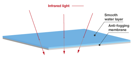
cap-ONE uses an original transparent film with an anti-fogging membrane. It forms a smooth layer of water which allows stable transmission of infrared light without irregular rejection. This technology eliminates the need for heaters and reduces the power consumption and weight of the sensor.


Without anti-fogging membrane

With anti-fogging membrane inside the airway
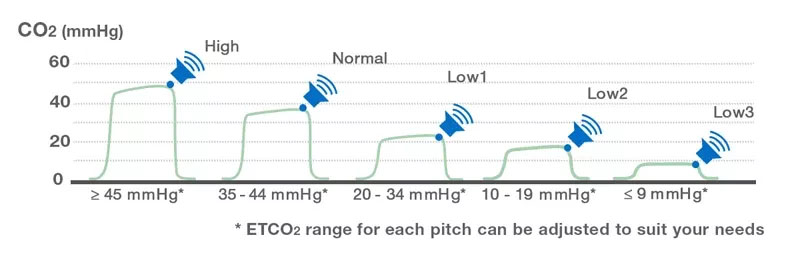
Image
image tech co2 05
Image
image tech co2 06
Image
image tech co2 07
Image
image tech co2 08
Improving manual ventilation and CPR with ETCO2 Audible Cue
Image
logo audible cue
Nihon Kohden developed ETCO2 Audible Cue that could monitor a patient’s respiratory status in real time by applying the technology we have cultivated through the development of mainstream CO2 sensor. ETCO2 Audible Cue helps the caregiver to manage ETCO2 during manual ventilation and CPR by delivering five different, easy recognizable sounds to indicate five ranges (Figure 1) from high to low so that they can determine the patient’s condition without looking at the monitor.
Image
Image tech cap-ONE 10
Figure 1. Audible Cue -Five ranges of sounds-
The actual Audible Cue sounds
Primarily designed forETCO2 RangeSound TypeSound DesignExample of SituationSound
Manual ventilation and ROSC confirmationHigh          
45≤
Image
Technology CO2 image 11
Image
Technology CO2 image 16
  • Hypoventilation is caused by insufficient manual ventilation during transport
  • ROSC is achieved*
Normal          
35-44
Image
Technology CO2 image 12
Image
Technology CO2 image 17
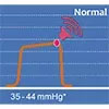
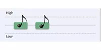
Ventilation is sufficient
Low1          
20-34
Image
Technology CO2 image 13
Image
Technology CO2 image 18
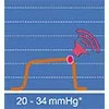
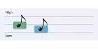
Hyperventilation is caused by over aggressive manual ventilation during transport
CPR            
(Chest compression)
Low2            
10-19
Image
Technology CO2 image 14
Image
Technology CO2 image 19
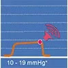
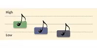
High quality chest compressions are achieved
Low3            
≤9
Image
Technology CO2 image 15
Image
Technology CO2 image 20
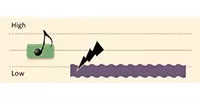
Quality of effectiveness of chest compression may need improvement

ROSC: Return of Spontaneous Circulation

Material Downloads - cap-ONE

Learn more
Image
thumb tech cap-one pdf 01

Monitor Your Patient with Reliable cap-ONE Mainstream CO2 Sensor

Image
thumb tech cap-one pdf 02

Ensure Quality of Care under Sedation

Image
thumb tech cap-one pdf 03

Optimal respiratory monitoring during sedation for all patients

Image
thumb tech cap-one pdf 04

Improve Manual Ventilation and CPR with ETCO2 Audible Cue

Publications

Video